Zinc in PDB 6ksn: Structure of A Zn-Bound Camelid Single Domain Antibody
Protein crystallography data
The structure of Structure of A Zn-Bound Camelid Single Domain Antibody, PDB code: 6ksn
was solved by
S.Kumar,
A.Athreya,
A.Penmatsa,
with X-Ray Crystallography technique. A brief refinement statistics is given in the table below:
| Resolution Low / High (Å) | 46.28 / 2.15 |
| Space group | P 32 2 1 |
| Cell size a, b, c (Å), α, β, γ (°) | 106.880, 106.880, 70.580, 90.00, 90.00, 120.00 |
| R / Rfree (%) | 19.3 / 23.2 |
Other elements in 6ksn:
The structure of Structure of A Zn-Bound Camelid Single Domain Antibody also contains other interesting chemical elements:
| Sodium | (Na) | 1 atom |
Zinc Binding Sites:
The binding sites of Zinc atom in the Structure of A Zn-Bound Camelid Single Domain Antibody
(pdb code 6ksn). This binding sites where shown within
5.0 Angstroms radius around Zinc atom.
In total 2 binding sites of Zinc where determined in the Structure of A Zn-Bound Camelid Single Domain Antibody, PDB code: 6ksn:
Jump to Zinc binding site number: 1; 2;
In total 2 binding sites of Zinc where determined in the Structure of A Zn-Bound Camelid Single Domain Antibody, PDB code: 6ksn:
Jump to Zinc binding site number: 1; 2;
Zinc binding site 1 out of 2 in 6ksn
Go back to
Zinc binding site 1 out
of 2 in the Structure of A Zn-Bound Camelid Single Domain Antibody
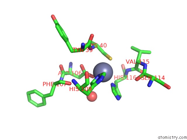
Mono view
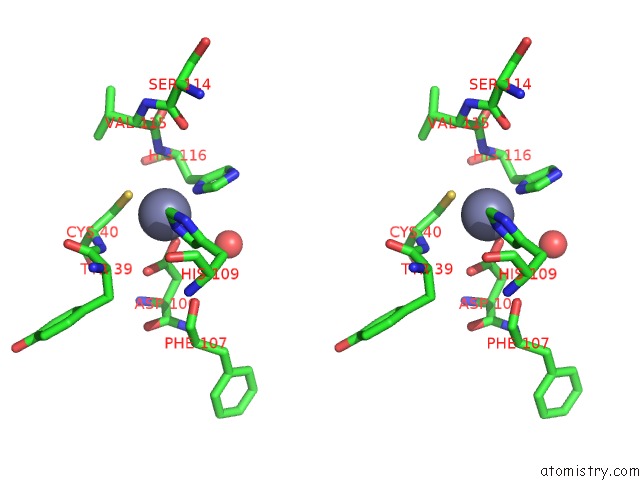
Stereo pair view
Mono view
Stereo pair view
A full contact list of Zinc with other atoms in the Zn binding
site number 1 of Structure of A Zn-Bound Camelid Single Domain Antibody within 5.0Å range:
|
Zinc binding site 2 out of 2 in 6ksn
Go back to
Zinc binding site 2 out
of 2 in the Structure of A Zn-Bound Camelid Single Domain Antibody
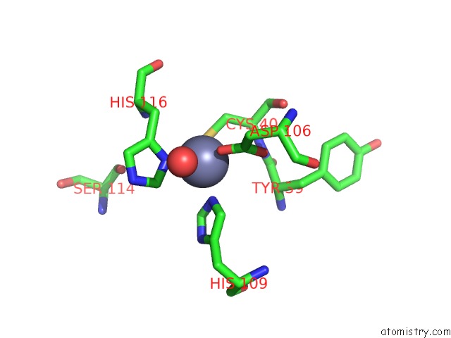
Mono view
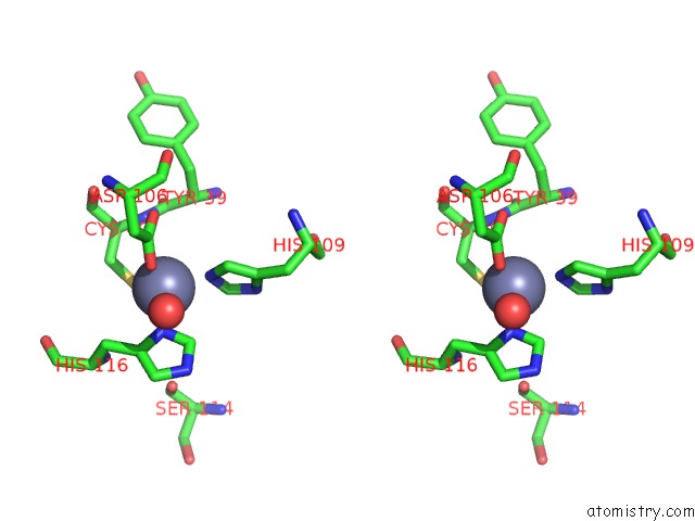
Stereo pair view
Mono view
Stereo pair view
A full contact list of Zinc with other atoms in the Zn binding
site number 2 of Structure of A Zn-Bound Camelid Single Domain Antibody within 5.0Å range:
|
Reference:
S.Kumar,
M.Ithayaraja,
A.Athreya,
R.Ranjan,
A.Penmatsa.
Isolation and Structural Characterization of A ZN2+-Bound Single-Domain Antibody Against Norc, A Putative Multi-Drug Efflux Transporter in Bacteria. J.Biol.Chem. 2019.
ISSN: ESSN 1083-351X
PubMed: 31699895
DOI: 10.1074/JBC.RA119.010902
Page generated: Tue Oct 29 02:06:17 2024
ISSN: ESSN 1083-351X
PubMed: 31699895
DOI: 10.1074/JBC.RA119.010902
Last articles
Zn in 9J0NZn in 9J0O
Zn in 9J0P
Zn in 9FJX
Zn in 9EKB
Zn in 9C0F
Zn in 9CAH
Zn in 9CH0
Zn in 9CH3
Zn in 9CH1