Zinc in PDB 5mcv: New Insights Into the Role of Dna Shape on Its Recognition By P53 Proteins (Complex P53DBD-LWC1)
Protein crystallography data
The structure of New Insights Into the Role of Dna Shape on Its Recognition By P53 Proteins (Complex P53DBD-LWC1), PDB code: 5mcv
was solved by
D.Golovenko,
H.Rozenberg,
Z.Shakked,
with X-Ray Crystallography technique. A brief refinement statistics is given in the table below:
| Resolution Low / High (Å) | 34.48 / 1.60 |
| Space group | C 1 2 1 |
| Cell size a, b, c (Å), α, β, γ (°) | 138.078, 49.533, 68.047, 90.00, 92.91, 90.00 |
| R / Rfree (%) | 15.4 / 18.8 |
Zinc Binding Sites:
The binding sites of Zinc atom in the New Insights Into the Role of Dna Shape on Its Recognition By P53 Proteins (Complex P53DBD-LWC1)
(pdb code 5mcv). This binding sites where shown within
5.0 Angstroms radius around Zinc atom.
In total 2 binding sites of Zinc where determined in the New Insights Into the Role of Dna Shape on Its Recognition By P53 Proteins (Complex P53DBD-LWC1), PDB code: 5mcv:
Jump to Zinc binding site number: 1; 2;
In total 2 binding sites of Zinc where determined in the New Insights Into the Role of Dna Shape on Its Recognition By P53 Proteins (Complex P53DBD-LWC1), PDB code: 5mcv:
Jump to Zinc binding site number: 1; 2;
Zinc binding site 1 out of 2 in 5mcv
Go back to
Zinc binding site 1 out
of 2 in the New Insights Into the Role of Dna Shape on Its Recognition By P53 Proteins (Complex P53DBD-LWC1)
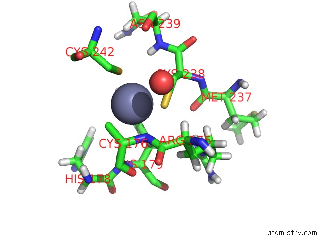
Mono view
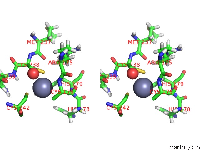
Stereo pair view
Mono view
Stereo pair view
A full contact list of Zinc with other atoms in the Zn binding
site number 1 of New Insights Into the Role of Dna Shape on Its Recognition By P53 Proteins (Complex P53DBD-LWC1) within 5.0Å range:
|
Zinc binding site 2 out of 2 in 5mcv
Go back to
Zinc binding site 2 out
of 2 in the New Insights Into the Role of Dna Shape on Its Recognition By P53 Proteins (Complex P53DBD-LWC1)
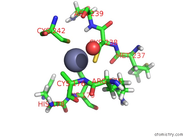
Mono view
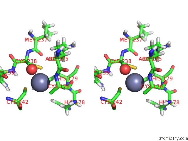
Stereo pair view
Mono view
Stereo pair view
A full contact list of Zinc with other atoms in the Zn binding
site number 2 of New Insights Into the Role of Dna Shape on Its Recognition By P53 Proteins (Complex P53DBD-LWC1) within 5.0Å range:
|
Reference:
D.Golovenko,
B.Brauning,
P.Vyas,
T.E.Haran,
H.Rozenberg,
Z.Shakked.
New Insights Into the Role of Dna Shape on Its Recognition By P53 Proteins. Structure V. 26 1237 2018.
ISSN: ISSN 1878-4186
PubMed: 30057026
DOI: 10.1016/J.STR.2018.06.006
Page generated: Sun Oct 27 22:06:13 2024
ISSN: ISSN 1878-4186
PubMed: 30057026
DOI: 10.1016/J.STR.2018.06.006
Last articles
Zn in 9J0NZn in 9J0O
Zn in 9J0P
Zn in 9FJX
Zn in 9EKB
Zn in 9C0F
Zn in 9CAH
Zn in 9CH0
Zn in 9CH3
Zn in 9CH1