Zinc in PDB 5kiv: Crystal Structure of Saumacro (SAV0325)
Protein crystallography data
The structure of Crystal Structure of Saumacro (SAV0325), PDB code: 5kiv
was solved by
R.S.Williams,
C.D.Appel,
G.K.Feld,
B.D.Wallace,
with X-Ray Crystallography technique. A brief refinement statistics is given in the table below:
| Resolution Low / High (Å) | 29.98 / 1.75 |
| Space group | P 21 21 21 |
| Cell size a, b, c (Å), α, β, γ (°) | 46.823, 47.853, 134.950, 90.00, 90.00, 90.00 |
| R / Rfree (%) | 15.7 / 19.4 |
Zinc Binding Sites:
The binding sites of Zinc atom in the Crystal Structure of Saumacro (SAV0325)
(pdb code 5kiv). This binding sites where shown within
5.0 Angstroms radius around Zinc atom.
In total only one binding site of Zinc was determined in the Crystal Structure of Saumacro (SAV0325), PDB code: 5kiv:
In total only one binding site of Zinc was determined in the Crystal Structure of Saumacro (SAV0325), PDB code: 5kiv:
Zinc binding site 1 out of 1 in 5kiv
Go back to
Zinc binding site 1 out
of 1 in the Crystal Structure of Saumacro (SAV0325)
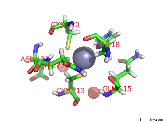
Mono view
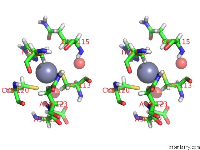
Stereo pair view
Mono view
Stereo pair view
A full contact list of Zinc with other atoms in the Zn binding
site number 1 of Crystal Structure of Saumacro (SAV0325) within 5.0Å range:
|
Reference:
C.D.Appel,
G.K.Feld,
B.D.Wallace,
R.S.Williams.
Structure of the Sirtuin-Linked Macrodomain SAV0325 From Staphylococcus Aureus. Protein Sci. V. 25 1682 2016.
ISSN: ESSN 1469-896X
PubMed: 27345688
DOI: 10.1002/PRO.2974
Page generated: Sun Oct 27 20:27:07 2024
ISSN: ESSN 1469-896X
PubMed: 27345688
DOI: 10.1002/PRO.2974
Last articles
Zn in 9J0NZn in 9J0O
Zn in 9J0P
Zn in 9FJX
Zn in 9EKB
Zn in 9C0F
Zn in 9CAH
Zn in 9CH0
Zn in 9CH3
Zn in 9CH1