Zinc in PDB 5ab9: Structure of the P53 Cancer Mutant Y220C with Bound Small Molecule 7- Ethyl-3-(Piperidin-4-Yl)-1H-Indole
Protein crystallography data
The structure of Structure of the P53 Cancer Mutant Y220C with Bound Small Molecule 7- Ethyl-3-(Piperidin-4-Yl)-1H-Indole, PDB code: 5ab9
was solved by
A.C.Joerger,
with X-Ray Crystallography technique. A brief refinement statistics is given in the table below:
| Resolution Low / High (Å) | 29.43 / 1.36 |
| Space group | P 21 21 21 |
| Cell size a, b, c (Å), α, β, γ (°) | 64.994, 71.059, 105.028, 90.00, 90.00, 90.00 |
| R / Rfree (%) | 14.3 / 16.6 |
Zinc Binding Sites:
The binding sites of Zinc atom in the Structure of the P53 Cancer Mutant Y220C with Bound Small Molecule 7- Ethyl-3-(Piperidin-4-Yl)-1H-Indole
(pdb code 5ab9). This binding sites where shown within
5.0 Angstroms radius around Zinc atom.
In total 2 binding sites of Zinc where determined in the Structure of the P53 Cancer Mutant Y220C with Bound Small Molecule 7- Ethyl-3-(Piperidin-4-Yl)-1H-Indole, PDB code: 5ab9:
Jump to Zinc binding site number: 1; 2;
In total 2 binding sites of Zinc where determined in the Structure of the P53 Cancer Mutant Y220C with Bound Small Molecule 7- Ethyl-3-(Piperidin-4-Yl)-1H-Indole, PDB code: 5ab9:
Jump to Zinc binding site number: 1; 2;
Zinc binding site 1 out of 2 in 5ab9
Go back to
Zinc binding site 1 out
of 2 in the Structure of the P53 Cancer Mutant Y220C with Bound Small Molecule 7- Ethyl-3-(Piperidin-4-Yl)-1H-Indole
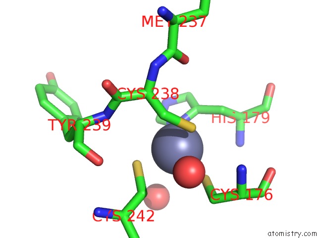
Mono view
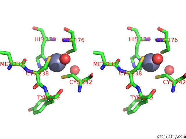
Stereo pair view
Mono view
Stereo pair view
A full contact list of Zinc with other atoms in the Zn binding
site number 1 of Structure of the P53 Cancer Mutant Y220C with Bound Small Molecule 7- Ethyl-3-(Piperidin-4-Yl)-1H-Indole within 5.0Å range:
|
Zinc binding site 2 out of 2 in 5ab9
Go back to
Zinc binding site 2 out
of 2 in the Structure of the P53 Cancer Mutant Y220C with Bound Small Molecule 7- Ethyl-3-(Piperidin-4-Yl)-1H-Indole
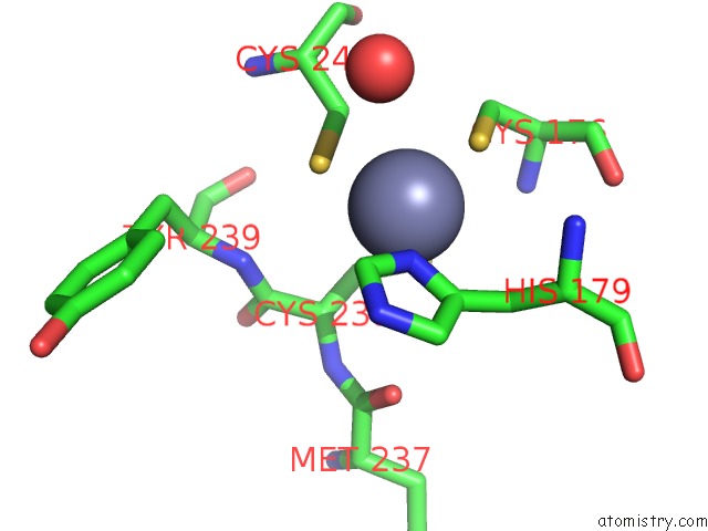
Mono view
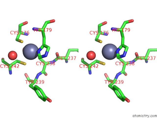
Stereo pair view
Mono view
Stereo pair view
A full contact list of Zinc with other atoms in the Zn binding
site number 2 of Structure of the P53 Cancer Mutant Y220C with Bound Small Molecule 7- Ethyl-3-(Piperidin-4-Yl)-1H-Indole within 5.0Å range:
|
Reference:
A.C.Joerger,
M.R.Bauer,
R.Wilcken,
M.G.J.Baud,
H.Harbrecht,
T.E.Exner,
F.M.Boeckler,
J.Spencer,
A.R.Fersht.
Exploiting Transient Protein States For the Design of Small-Molecule Stabilizers of Mutant P53. Structure V. 23 2246 2015.
ISSN: ISSN 0969-2126
PubMed: 26636255
DOI: 10.1016/J.STR.2015.10.016
Page generated: Sun Oct 27 12:47:10 2024
ISSN: ISSN 0969-2126
PubMed: 26636255
DOI: 10.1016/J.STR.2015.10.016
Last articles
Zn in 9J0NZn in 9J0O
Zn in 9J0P
Zn in 9FJX
Zn in 9EKB
Zn in 9C0F
Zn in 9CAH
Zn in 9CH0
Zn in 9CH3
Zn in 9CH1