Zinc in PDB 4q3j: Crystal Structure of Nfkb-P65-Degrading Zinc Protease Family Protein
Protein crystallography data
The structure of Crystal Structure of Nfkb-P65-Degrading Zinc Protease Family Protein, PDB code: 4q3j
was solved by
M.C.Sousa,
M.M.Turco,
with X-Ray Crystallography technique. A brief refinement statistics is given in the table below:
| Resolution Low / High (Å) | 34.94 / 1.86 |
| Space group | P 21 21 21 |
| Cell size a, b, c (Å), α, β, γ (°) | 45.205, 67.480, 81.688, 90.00, 90.00, 90.00 |
| R / Rfree (%) | 17.9 / 21.8 |
Other elements in 4q3j:
The structure of Crystal Structure of Nfkb-P65-Degrading Zinc Protease Family Protein also contains other interesting chemical elements:
| Magnesium | (Mg) | 1 atom |
Zinc Binding Sites:
The binding sites of Zinc atom in the Crystal Structure of Nfkb-P65-Degrading Zinc Protease Family Protein
(pdb code 4q3j). This binding sites where shown within
5.0 Angstroms radius around Zinc atom.
In total only one binding site of Zinc was determined in the Crystal Structure of Nfkb-P65-Degrading Zinc Protease Family Protein, PDB code: 4q3j:
In total only one binding site of Zinc was determined in the Crystal Structure of Nfkb-P65-Degrading Zinc Protease Family Protein, PDB code: 4q3j:
Zinc binding site 1 out of 1 in 4q3j
Go back to
Zinc binding site 1 out
of 1 in the Crystal Structure of Nfkb-P65-Degrading Zinc Protease Family Protein
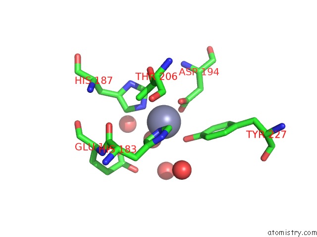
Mono view
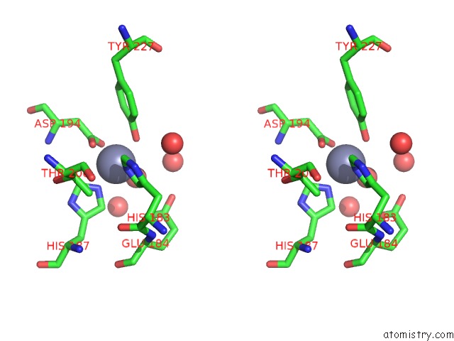
Stereo pair view
Mono view
Stereo pair view
A full contact list of Zinc with other atoms in the Zn binding
site number 1 of Crystal Structure of Nfkb-P65-Degrading Zinc Protease Family Protein within 5.0Å range:
|
Reference:
M.M.Turco,
M.C.Sousa.
The Structure and Specificity of the Type III Secretion System Effector Nlec Suggest A Dna Mimicry Mechanism of Substrate Recognition. Biochemistry V. 53 5131 2014.
ISSN: ISSN 0006-2960
PubMed: 25040221
DOI: 10.1021/BI500593E
Page generated: Sun Oct 27 06:15:02 2024
ISSN: ISSN 0006-2960
PubMed: 25040221
DOI: 10.1021/BI500593E
Last articles
Zn in 9J0NZn in 9J0O
Zn in 9J0P
Zn in 9FJX
Zn in 9EKB
Zn in 9C0F
Zn in 9CAH
Zn in 9CH0
Zn in 9CH3
Zn in 9CH1