Zinc in PDB 4o6s: 1.32A Resolution Structure of the Hemophore Hasa From Pseudomonas Aeruginosa (H83A Mutant, Zinc Bound)
Protein crystallography data
The structure of 1.32A Resolution Structure of the Hemophore Hasa From Pseudomonas Aeruginosa (H83A Mutant, Zinc Bound), PDB code: 4o6s
was solved by
S.Lovell,
R.Kumar,
K.P.Battaile,
H.Matsumura,
H.Yao,
J.C.Rodriguez,
P.Moenne-Loccoz,
M.Rivera,
with X-Ray Crystallography technique. A brief refinement statistics is given in the table below:
| Resolution Low / High (Å) | 34.63 / 1.32 |
| Space group | P 1 21 1 |
| Cell size a, b, c (Å), α, β, γ (°) | 34.898, 66.232, 40.999, 90.00, 97.13, 90.00 |
| R / Rfree (%) | 14.3 / 16.8 |
Other elements in 4o6s:
The structure of 1.32A Resolution Structure of the Hemophore Hasa From Pseudomonas Aeruginosa (H83A Mutant, Zinc Bound) also contains other interesting chemical elements:
| Iron | (Fe) | 1 atom |
Zinc Binding Sites:
The binding sites of Zinc atom in the 1.32A Resolution Structure of the Hemophore Hasa From Pseudomonas Aeruginosa (H83A Mutant, Zinc Bound)
(pdb code 4o6s). This binding sites where shown within
5.0 Angstroms radius around Zinc atom.
In total 9 binding sites of Zinc where determined in the 1.32A Resolution Structure of the Hemophore Hasa From Pseudomonas Aeruginosa (H83A Mutant, Zinc Bound), PDB code: 4o6s:
Jump to Zinc binding site number: 1; 2; 3; 4; 5; 6; 7; 8; 9;
In total 9 binding sites of Zinc where determined in the 1.32A Resolution Structure of the Hemophore Hasa From Pseudomonas Aeruginosa (H83A Mutant, Zinc Bound), PDB code: 4o6s:
Jump to Zinc binding site number: 1; 2; 3; 4; 5; 6; 7; 8; 9;
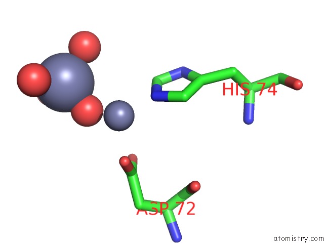
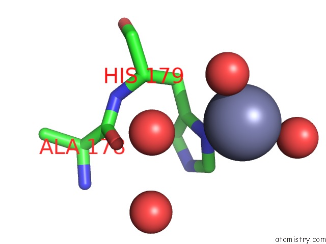
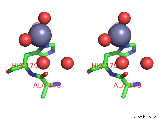
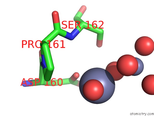
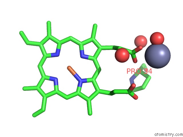
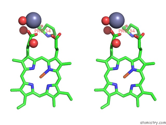
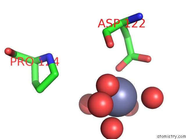
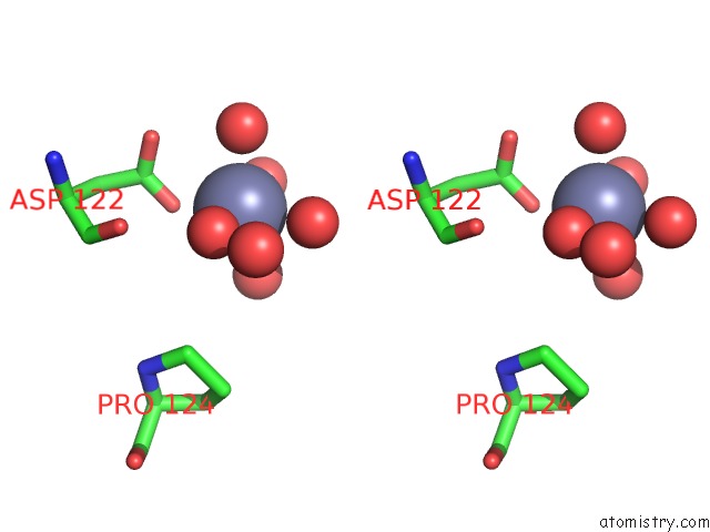
Zinc binding site 1 out of 9 in 4o6s
Go back to
Zinc binding site 1 out
of 9 in the 1.32A Resolution Structure of the Hemophore Hasa From Pseudomonas Aeruginosa (H83A Mutant, Zinc Bound)
Mono view
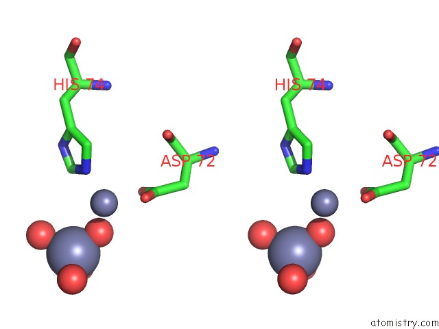
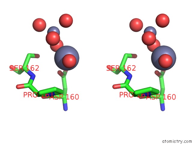
Stereo pair view
Mono view
Stereo pair view
A full contact list of Zinc with other atoms in the Zn binding
site number 1 of 1.32A Resolution Structure of the Hemophore Hasa From Pseudomonas Aeruginosa (H83A Mutant, Zinc Bound) within 5.0Å range:
|
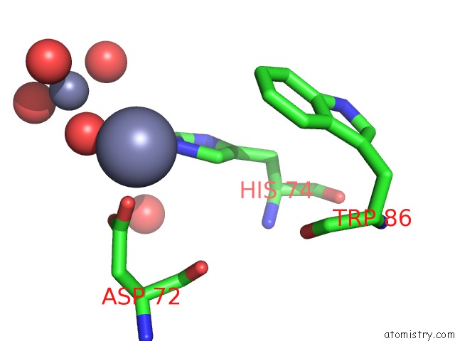
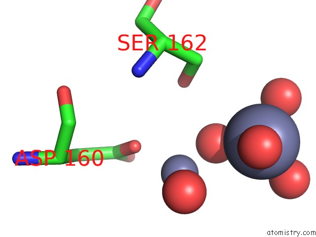
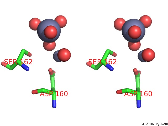
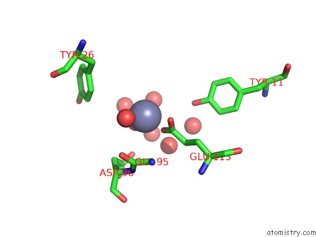
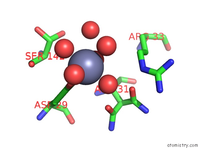
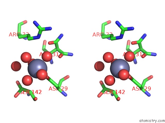
Zinc binding site 2 out of 9 in 4o6s
Go back to
Zinc binding site 2 out
of 9 in the 1.32A Resolution Structure of the Hemophore Hasa From Pseudomonas Aeruginosa (H83A Mutant, Zinc Bound)
Mono view
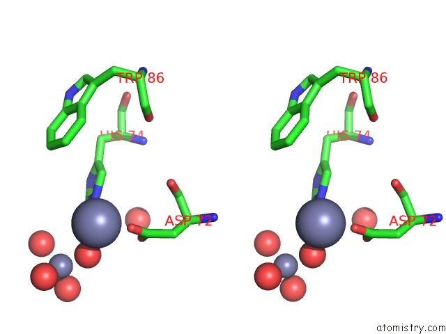
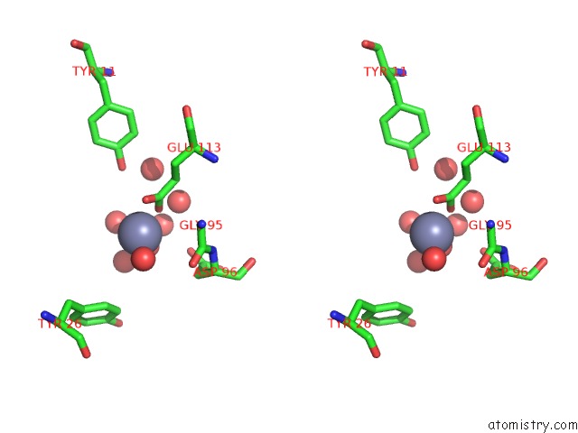
Stereo pair view
Mono view
Stereo pair view
A full contact list of Zinc with other atoms in the Zn binding
site number 2 of 1.32A Resolution Structure of the Hemophore Hasa From Pseudomonas Aeruginosa (H83A Mutant, Zinc Bound) within 5.0Å range:
|
Zinc binding site 3 out of 9 in 4o6s
Go back to
Zinc binding site 3 out
of 9 in the 1.32A Resolution Structure of the Hemophore Hasa From Pseudomonas Aeruginosa (H83A Mutant, Zinc Bound)
Mono view
Stereo pair view
Mono view
Stereo pair view
A full contact list of Zinc with other atoms in the Zn binding
site number 3 of 1.32A Resolution Structure of the Hemophore Hasa From Pseudomonas Aeruginosa (H83A Mutant, Zinc Bound) within 5.0Å range:
|
Zinc binding site 4 out of 9 in 4o6s
Go back to
Zinc binding site 4 out
of 9 in the 1.32A Resolution Structure of the Hemophore Hasa From Pseudomonas Aeruginosa (H83A Mutant, Zinc Bound)
Mono view
Stereo pair view
Mono view
Stereo pair view
A full contact list of Zinc with other atoms in the Zn binding
site number 4 of 1.32A Resolution Structure of the Hemophore Hasa From Pseudomonas Aeruginosa (H83A Mutant, Zinc Bound) within 5.0Å range:
|
Zinc binding site 5 out of 9 in 4o6s
Go back to
Zinc binding site 5 out
of 9 in the 1.32A Resolution Structure of the Hemophore Hasa From Pseudomonas Aeruginosa (H83A Mutant, Zinc Bound)
Mono view
Stereo pair view
Mono view
Stereo pair view
A full contact list of Zinc with other atoms in the Zn binding
site number 5 of 1.32A Resolution Structure of the Hemophore Hasa From Pseudomonas Aeruginosa (H83A Mutant, Zinc Bound) within 5.0Å range:
|
Zinc binding site 6 out of 9 in 4o6s
Go back to
Zinc binding site 6 out
of 9 in the 1.32A Resolution Structure of the Hemophore Hasa From Pseudomonas Aeruginosa (H83A Mutant, Zinc Bound)
Mono view
Stereo pair view
Mono view
Stereo pair view
A full contact list of Zinc with other atoms in the Zn binding
site number 6 of 1.32A Resolution Structure of the Hemophore Hasa From Pseudomonas Aeruginosa (H83A Mutant, Zinc Bound) within 5.0Å range:
|
Zinc binding site 7 out of 9 in 4o6s
Go back to
Zinc binding site 7 out
of 9 in the 1.32A Resolution Structure of the Hemophore Hasa From Pseudomonas Aeruginosa (H83A Mutant, Zinc Bound)
Mono view
Stereo pair view
Mono view
Stereo pair view
A full contact list of Zinc with other atoms in the Zn binding
site number 7 of 1.32A Resolution Structure of the Hemophore Hasa From Pseudomonas Aeruginosa (H83A Mutant, Zinc Bound) within 5.0Å range:
|
Zinc binding site 8 out of 9 in 4o6s
Go back to
Zinc binding site 8 out
of 9 in the 1.32A Resolution Structure of the Hemophore Hasa From Pseudomonas Aeruginosa (H83A Mutant, Zinc Bound)
Mono view
Stereo pair view
Mono view
Stereo pair view
A full contact list of Zinc with other atoms in the Zn binding
site number 8 of 1.32A Resolution Structure of the Hemophore Hasa From Pseudomonas Aeruginosa (H83A Mutant, Zinc Bound) within 5.0Å range:
|
Zinc binding site 9 out of 9 in 4o6s
Go back to
Zinc binding site 9 out
of 9 in the 1.32A Resolution Structure of the Hemophore Hasa From Pseudomonas Aeruginosa (H83A Mutant, Zinc Bound)
Mono view
Stereo pair view
Mono view
Stereo pair view
A full contact list of Zinc with other atoms in the Zn binding
site number 9 of 1.32A Resolution Structure of the Hemophore Hasa From Pseudomonas Aeruginosa (H83A Mutant, Zinc Bound) within 5.0Å range:
|
Reference:
R.Kumar,
H.Matsumura,
S.Lovell,
H.Yao,
J.C.Rodriguez,
K.P.Battaile,
P.Moenne-Loccoz,
M.Rivera.
Replacing the Axial Ligand Tyrosine 75 or Its Hydrogen Bond Partner Histidine 83 Minimally Affects Hemin Acquisition By the Hemophore Hasap From Pseudomonas Aeruginosa. Biochemistry V. 53 2112 2014.
ISSN: ISSN 0006-2960
PubMed: 24625274
DOI: 10.1021/BI500030P
Page generated: Sun Oct 27 03:37:04 2024
ISSN: ISSN 0006-2960
PubMed: 24625274
DOI: 10.1021/BI500030P
Last articles
Zn in 9J0NZn in 9J0O
Zn in 9J0P
Zn in 9FJX
Zn in 9EKB
Zn in 9C0F
Zn in 9CAH
Zn in 9CH0
Zn in 9CH3
Zn in 9CH1