Zinc in PDB 4i7d: SIAH1 Bound to Synthetic Peptide (Ace)Klrpvamvrp(Prk)Vr
Protein crystallography data
The structure of SIAH1 Bound to Synthetic Peptide (Ace)Klrpvamvrp(Prk)Vr, PDB code: 4i7d
was solved by
E.Santelli,
J.L.Stebbins,
Y.Feng,
S.K.De,
A.Purves,
K.Motamedchaboki,
B.Wu,
Z.A.Ronai,
R.C.Liddington,
M.Pellecchia,
with X-Ray Crystallography technique. A brief refinement statistics is given in the table below:
| Resolution Low / High (Å) | 30.00 / 2.40 |
| Space group | I 2 3 |
| Cell size a, b, c (Å), α, β, γ (°) | 160.977, 160.977, 160.977, 90.00, 90.00, 90.00 |
| R / Rfree (%) | 17.1 / 20.2 |
Zinc Binding Sites:
The binding sites of Zinc atom in the SIAH1 Bound to Synthetic Peptide (Ace)Klrpvamvrp(Prk)Vr
(pdb code 4i7d). This binding sites where shown within
5.0 Angstroms radius around Zinc atom.
In total 4 binding sites of Zinc where determined in the SIAH1 Bound to Synthetic Peptide (Ace)Klrpvamvrp(Prk)Vr, PDB code: 4i7d:
Jump to Zinc binding site number: 1; 2; 3; 4;
In total 4 binding sites of Zinc where determined in the SIAH1 Bound to Synthetic Peptide (Ace)Klrpvamvrp(Prk)Vr, PDB code: 4i7d:
Jump to Zinc binding site number: 1; 2; 3; 4;
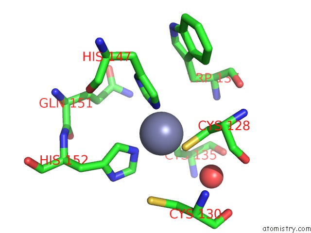
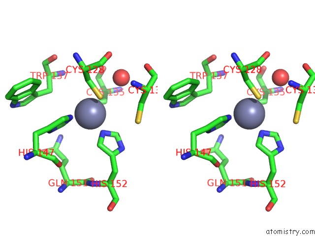
Zinc binding site 1 out of 4 in 4i7d
Go back to
Zinc binding site 1 out
of 4 in the SIAH1 Bound to Synthetic Peptide (Ace)Klrpvamvrp(Prk)Vr
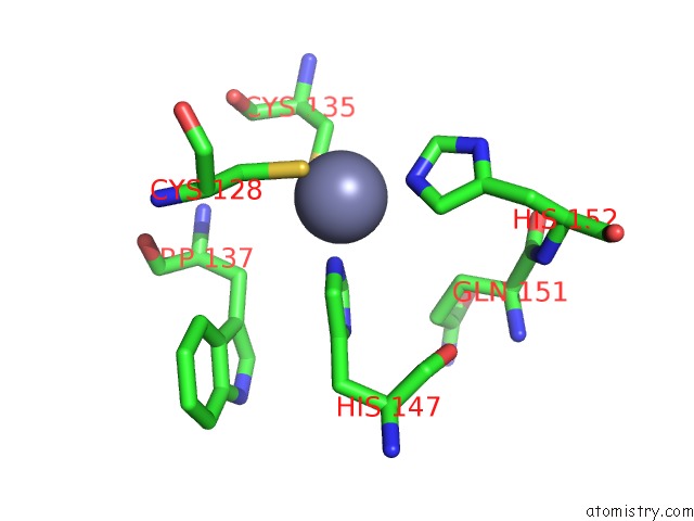
Mono view
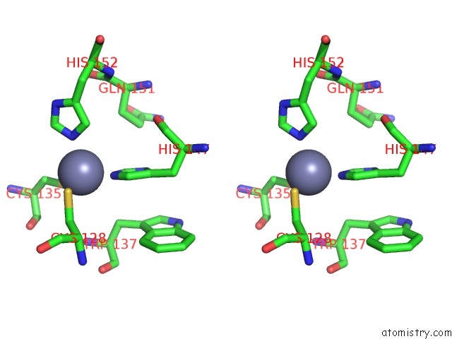
Stereo pair view
Mono view
Stereo pair view
A full contact list of Zinc with other atoms in the Zn binding
site number 1 of SIAH1 Bound to Synthetic Peptide (Ace)Klrpvamvrp(Prk)Vr within 5.0Å range:
|
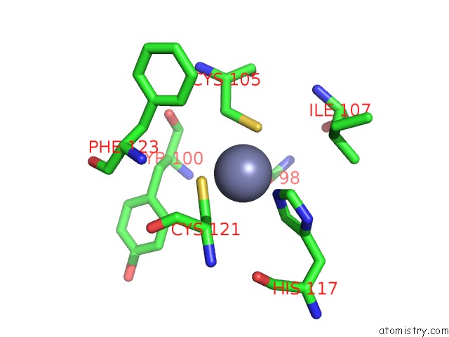
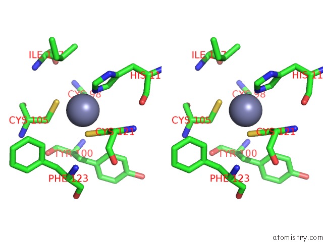
Zinc binding site 2 out of 4 in 4i7d
Go back to
Zinc binding site 2 out
of 4 in the SIAH1 Bound to Synthetic Peptide (Ace)Klrpvamvrp(Prk)Vr
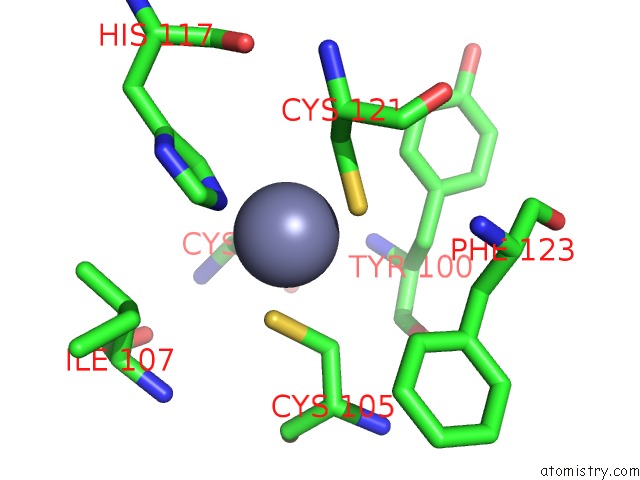
Mono view
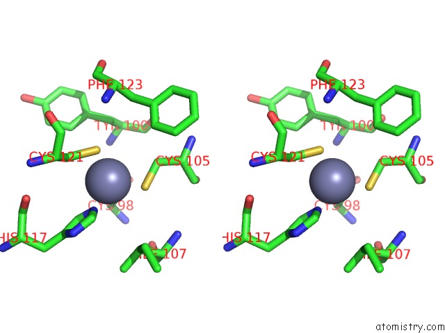
Stereo pair view
Mono view
Stereo pair view
A full contact list of Zinc with other atoms in the Zn binding
site number 2 of SIAH1 Bound to Synthetic Peptide (Ace)Klrpvamvrp(Prk)Vr within 5.0Å range:
|
Zinc binding site 3 out of 4 in 4i7d
Go back to
Zinc binding site 3 out
of 4 in the SIAH1 Bound to Synthetic Peptide (Ace)Klrpvamvrp(Prk)Vr
Mono view
Stereo pair view
Mono view
Stereo pair view
A full contact list of Zinc with other atoms in the Zn binding
site number 3 of SIAH1 Bound to Synthetic Peptide (Ace)Klrpvamvrp(Prk)Vr within 5.0Å range:
|
Zinc binding site 4 out of 4 in 4i7d
Go back to
Zinc binding site 4 out
of 4 in the SIAH1 Bound to Synthetic Peptide (Ace)Klrpvamvrp(Prk)Vr
Mono view
Stereo pair view
Mono view
Stereo pair view
A full contact list of Zinc with other atoms in the Zn binding
site number 4 of SIAH1 Bound to Synthetic Peptide (Ace)Klrpvamvrp(Prk)Vr within 5.0Å range:
|
Reference:
J.L.Stebbins,
E.Santelli,
Y.Feng,
S.K.De,
A.Purves,
K.Motamedchaboki,
B.Wu,
Z.A.Ronai,
R.C.Liddington,
M.Pellecchia.
Structure-Based Design of Covalent Siah Inhibitors. Chem.Biol. V. 20 973 2013.
ISSN: ISSN 1074-5521
PubMed: 23891150
DOI: 10.1016/J.CHEMBIOL.2013.06.008
Page generated: Sun Oct 27 00:35:07 2024
ISSN: ISSN 1074-5521
PubMed: 23891150
DOI: 10.1016/J.CHEMBIOL.2013.06.008
Last articles
Zn in 9J0NZn in 9J0O
Zn in 9J0P
Zn in 9FJX
Zn in 9EKB
Zn in 9C0F
Zn in 9CAH
Zn in 9CH0
Zn in 9CH3
Zn in 9CH1