Zinc in PDB 4c6n: Crystal Structure of the Dihydroorotase Domain of Human Cad E1637T Mutant Bound to Substrate at pH 6.0
Enzymatic activity of Crystal Structure of the Dihydroorotase Domain of Human Cad E1637T Mutant Bound to Substrate at pH 6.0
All present enzymatic activity of Crystal Structure of the Dihydroorotase Domain of Human Cad E1637T Mutant Bound to Substrate at pH 6.0:
3.5.2.3;
3.5.2.3;
Protein crystallography data
The structure of Crystal Structure of the Dihydroorotase Domain of Human Cad E1637T Mutant Bound to Substrate at pH 6.0, PDB code: 4c6n
was solved by
S.Ramon-Maiques,
N.Lallous,
A.Grande-Garcia,
with X-Ray Crystallography technique. A brief refinement statistics is given in the table below:
| Resolution Low / High (Å) | 48.399 / 1.90 |
| Space group | C 2 2 21 |
| Cell size a, b, c (Å), α, β, γ (°) | 82.100, 159.170, 60.970, 90.00, 90.00, 90.00 |
| R / Rfree (%) | 14.56 / 16.98 |
Zinc Binding Sites:
The binding sites of Zinc atom in the Crystal Structure of the Dihydroorotase Domain of Human Cad E1637T Mutant Bound to Substrate at pH 6.0
(pdb code 4c6n). This binding sites where shown within
5.0 Angstroms radius around Zinc atom.
In total 2 binding sites of Zinc where determined in the Crystal Structure of the Dihydroorotase Domain of Human Cad E1637T Mutant Bound to Substrate at pH 6.0, PDB code: 4c6n:
Jump to Zinc binding site number: 1; 2;
In total 2 binding sites of Zinc where determined in the Crystal Structure of the Dihydroorotase Domain of Human Cad E1637T Mutant Bound to Substrate at pH 6.0, PDB code: 4c6n:
Jump to Zinc binding site number: 1; 2;
Zinc binding site 1 out of 2 in 4c6n
Go back to
Zinc binding site 1 out
of 2 in the Crystal Structure of the Dihydroorotase Domain of Human Cad E1637T Mutant Bound to Substrate at pH 6.0
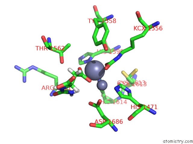
Mono view
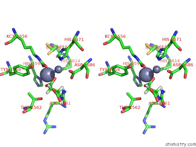
Stereo pair view
Mono view
Stereo pair view
A full contact list of Zinc with other atoms in the Zn binding
site number 1 of Crystal Structure of the Dihydroorotase Domain of Human Cad E1637T Mutant Bound to Substrate at pH 6.0 within 5.0Å range:
|
Zinc binding site 2 out of 2 in 4c6n
Go back to
Zinc binding site 2 out
of 2 in the Crystal Structure of the Dihydroorotase Domain of Human Cad E1637T Mutant Bound to Substrate at pH 6.0
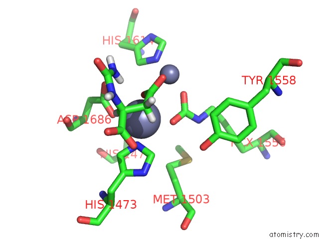
Mono view
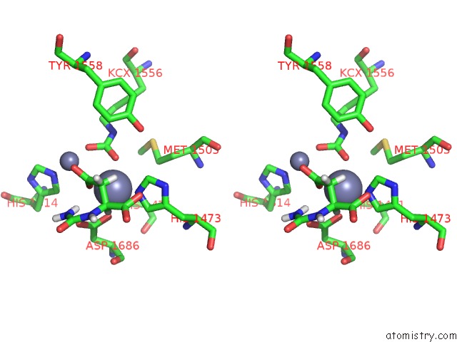
Stereo pair view
Mono view
Stereo pair view
A full contact list of Zinc with other atoms in the Zn binding
site number 2 of Crystal Structure of the Dihydroorotase Domain of Human Cad E1637T Mutant Bound to Substrate at pH 6.0 within 5.0Å range:
|
Reference:
A.Grande-Garcia,
N.Lallous,
C.Diaz-Tejada,
S.Ramon-Maiques.
Structure, Functional Characterization and Evolution of the Dihydroorotase Domain of Human Cad. Structure V. 22 185 2014.
ISSN: ISSN 0969-2126
PubMed: 24332717
DOI: 10.1016/J.STR.2013.10.016
Page generated: Sat Oct 26 20:38:42 2024
ISSN: ISSN 0969-2126
PubMed: 24332717
DOI: 10.1016/J.STR.2013.10.016
Last articles
Zn in 9J0NZn in 9J0O
Zn in 9J0P
Zn in 9FJX
Zn in 9EKB
Zn in 9C0F
Zn in 9CAH
Zn in 9CH0
Zn in 9CH3
Zn in 9CH1