Zinc in PDB 3sue: Crystal Structure of NS3/4A Protease Variant R155K in Complex with Mk- 5172
Protein crystallography data
The structure of Crystal Structure of NS3/4A Protease Variant R155K in Complex with Mk- 5172, PDB code: 3sue
was solved by
C.A.Schiffer,
K.P.Romano,
with X-Ray Crystallography technique. A brief refinement statistics is given in the table below:
| Resolution Low / High (Å) | 32.24 / 2.20 |
| Space group | P 1 21 1 |
| Cell size a, b, c (Å), α, β, γ (°) | 56.335, 103.324, 73.498, 90.00, 112.58, 90.00 |
| R / Rfree (%) | 18.6 / 22.9 |
Zinc Binding Sites:
The binding sites of Zinc atom in the Crystal Structure of NS3/4A Protease Variant R155K in Complex with Mk- 5172
(pdb code 3sue). This binding sites where shown within
5.0 Angstroms radius around Zinc atom.
In total 4 binding sites of Zinc where determined in the Crystal Structure of NS3/4A Protease Variant R155K in Complex with Mk- 5172, PDB code: 3sue:
Jump to Zinc binding site number: 1; 2; 3; 4;
In total 4 binding sites of Zinc where determined in the Crystal Structure of NS3/4A Protease Variant R155K in Complex with Mk- 5172, PDB code: 3sue:
Jump to Zinc binding site number: 1; 2; 3; 4;
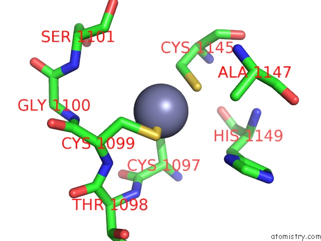
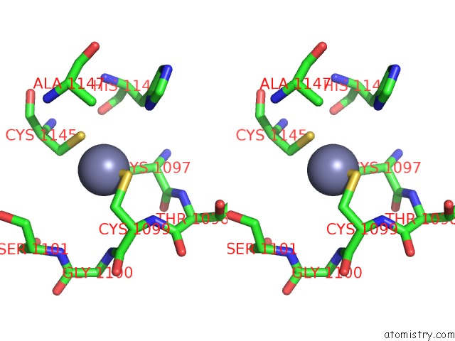
Zinc binding site 1 out of 4 in 3sue
Go back to
Zinc binding site 1 out
of 4 in the Crystal Structure of NS3/4A Protease Variant R155K in Complex with Mk- 5172
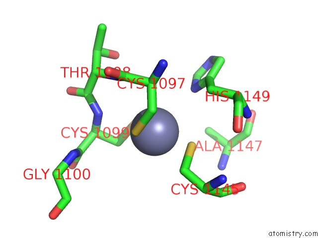
Mono view
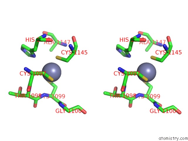
Stereo pair view
Mono view
Stereo pair view
A full contact list of Zinc with other atoms in the Zn binding
site number 1 of Crystal Structure of NS3/4A Protease Variant R155K in Complex with Mk- 5172 within 5.0Å range:
|
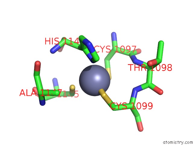
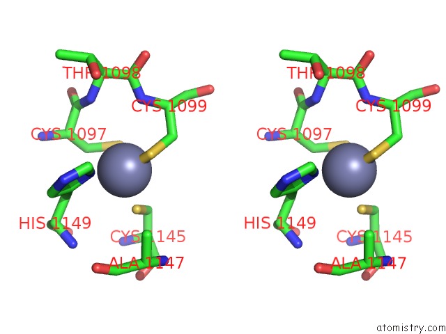
Zinc binding site 2 out of 4 in 3sue
Go back to
Zinc binding site 2 out
of 4 in the Crystal Structure of NS3/4A Protease Variant R155K in Complex with Mk- 5172
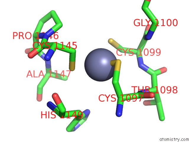
Mono view
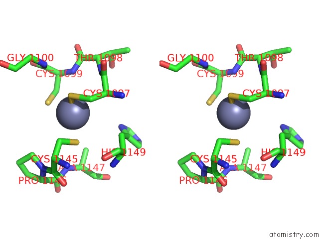
Stereo pair view
Mono view
Stereo pair view
A full contact list of Zinc with other atoms in the Zn binding
site number 2 of Crystal Structure of NS3/4A Protease Variant R155K in Complex with Mk- 5172 within 5.0Å range:
|
Zinc binding site 3 out of 4 in 3sue
Go back to
Zinc binding site 3 out
of 4 in the Crystal Structure of NS3/4A Protease Variant R155K in Complex with Mk- 5172
Mono view
Stereo pair view
Mono view
Stereo pair view
A full contact list of Zinc with other atoms in the Zn binding
site number 3 of Crystal Structure of NS3/4A Protease Variant R155K in Complex with Mk- 5172 within 5.0Å range:
|
Zinc binding site 4 out of 4 in 3sue
Go back to
Zinc binding site 4 out
of 4 in the Crystal Structure of NS3/4A Protease Variant R155K in Complex with Mk- 5172
Mono view
Stereo pair view
Mono view
Stereo pair view
A full contact list of Zinc with other atoms in the Zn binding
site number 4 of Crystal Structure of NS3/4A Protease Variant R155K in Complex with Mk- 5172 within 5.0Å range:
|
Reference:
K.P.Romano,
A.Ali,
C.Aydin,
D.Soumana,
A.Ozen,
L.M.Deveau,
C.Silver,
H.Cao,
A.Newton,
C.J.Petropoulos,
W.Huang,
C.A.Schiffer.
The Molecular Basis of Drug Resistance Against Hepatitis C Virus NS3/4A Protease Inhibitors. Plos Pathog. V. 8 02832 2012.
ISSN: ISSN 1553-7366
PubMed: 22910833
DOI: 10.1371/JOURNAL.PPAT.1002832
Page generated: Sat Oct 26 16:08:13 2024
ISSN: ISSN 1553-7366
PubMed: 22910833
DOI: 10.1371/JOURNAL.PPAT.1002832
Last articles
Zn in 9J0NZn in 9J0O
Zn in 9J0P
Zn in 9FJX
Zn in 9EKB
Zn in 9C0F
Zn in 9CAH
Zn in 9CH0
Zn in 9CH3
Zn in 9CH1