Zinc in PDB 2w14: High-Resolution Crystal Structure of the P-I Snake Venom Metalloproteinase BAP1 in Complex with A Peptidomimetic: Insights Into Inhibitor Binding
Protein crystallography data
The structure of High-Resolution Crystal Structure of the P-I Snake Venom Metalloproteinase BAP1 in Complex with A Peptidomimetic: Insights Into Inhibitor Binding, PDB code: 2w14
was solved by
T.J.Lingott,
C.Schleberger,
J.M.Gutierrez,
I.Merfort,
with X-Ray Crystallography technique. A brief refinement statistics is given in the table below:
| Resolution Low / High (Å) | 18.22 / 1.08 |
| Space group | P 21 21 21 |
| Cell size a, b, c (Å), α, β, γ (°) | 37.900, 59.800, 83.100, 90.00, 90.00, 90.00 |
| R / Rfree (%) | 11.8 / 14.3 |
Zinc Binding Sites:
The binding sites of Zinc atom in the High-Resolution Crystal Structure of the P-I Snake Venom Metalloproteinase BAP1 in Complex with A Peptidomimetic: Insights Into Inhibitor Binding
(pdb code 2w14). This binding sites where shown within
5.0 Angstroms radius around Zinc atom.
In total only one binding site of Zinc was determined in the High-Resolution Crystal Structure of the P-I Snake Venom Metalloproteinase BAP1 in Complex with A Peptidomimetic: Insights Into Inhibitor Binding, PDB code: 2w14:
In total only one binding site of Zinc was determined in the High-Resolution Crystal Structure of the P-I Snake Venom Metalloproteinase BAP1 in Complex with A Peptidomimetic: Insights Into Inhibitor Binding, PDB code: 2w14:
Zinc binding site 1 out of 1 in 2w14
Go back to
Zinc binding site 1 out
of 1 in the High-Resolution Crystal Structure of the P-I Snake Venom Metalloproteinase BAP1 in Complex with A Peptidomimetic: Insights Into Inhibitor Binding
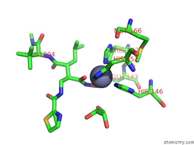
Mono view
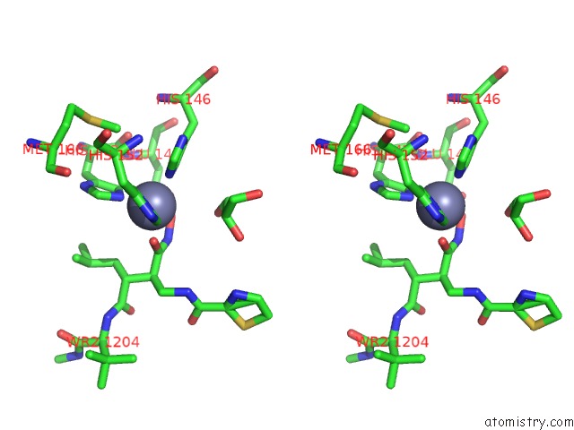
Stereo pair view
Mono view
Stereo pair view
A full contact list of Zinc with other atoms in the Zn binding
site number 1 of High-Resolution Crystal Structure of the P-I Snake Venom Metalloproteinase BAP1 in Complex with A Peptidomimetic: Insights Into Inhibitor Binding within 5.0Å range:
|
Reference:
T.J.Lingott,
C.Schleberger,
J.M.Gutierrez,
I.Merfort.
High-Resolution Crystal Structure of the Snake Venom Metalloproteinase BAP1 Complexed with A Peptidomimetic: Insight Into Inhibitor Binding. Biochemistry V. 48 6166 2009.
ISSN: ISSN 0006-2960
PubMed: 19485419
DOI: 10.1021/BI9002315
Page generated: Thu Oct 17 04:43:29 2024
ISSN: ISSN 0006-2960
PubMed: 19485419
DOI: 10.1021/BI9002315
Last articles
Zn in 9J0NZn in 9J0O
Zn in 9J0P
Zn in 9FJX
Zn in 9EKB
Zn in 9C0F
Zn in 9CAH
Zn in 9CH0
Zn in 9CH3
Zn in 9CH1