Zinc in PDB 1pv8: Crystal Structure of A Low Activity F12L Mutant of Human Porphobilinogen Synthase
Enzymatic activity of Crystal Structure of A Low Activity F12L Mutant of Human Porphobilinogen Synthase
All present enzymatic activity of Crystal Structure of A Low Activity F12L Mutant of Human Porphobilinogen Synthase:
4.2.1.24;
4.2.1.24;
Protein crystallography data
The structure of Crystal Structure of A Low Activity F12L Mutant of Human Porphobilinogen Synthase, PDB code: 1pv8
was solved by
S.Breinig,
J.Kervinen,
L.Stith,
A.S.Wasson,
R.Fairman,
A.Wlodawer,
A.Zdanov,
E.K.Jaffe,
with X-Ray Crystallography technique. A brief refinement statistics is given in the table below:
| Resolution Low / High (Å) | 45.00 / 2.20 |
| Space group | P 63 |
| Cell size a, b, c (Å), α, β, γ (°) | 89.571, 89.571, 153.190, 90.00, 90.00, 120.00 |
| R / Rfree (%) | 19.9 / 28.4 |
Zinc Binding Sites:
The binding sites of Zinc atom in the Crystal Structure of A Low Activity F12L Mutant of Human Porphobilinogen Synthase
(pdb code 1pv8). This binding sites where shown within
5.0 Angstroms radius around Zinc atom.
In total 2 binding sites of Zinc where determined in the Crystal Structure of A Low Activity F12L Mutant of Human Porphobilinogen Synthase, PDB code: 1pv8:
Jump to Zinc binding site number: 1; 2;
In total 2 binding sites of Zinc where determined in the Crystal Structure of A Low Activity F12L Mutant of Human Porphobilinogen Synthase, PDB code: 1pv8:
Jump to Zinc binding site number: 1; 2;
Zinc binding site 1 out of 2 in 1pv8
Go back to
Zinc binding site 1 out
of 2 in the Crystal Structure of A Low Activity F12L Mutant of Human Porphobilinogen Synthase
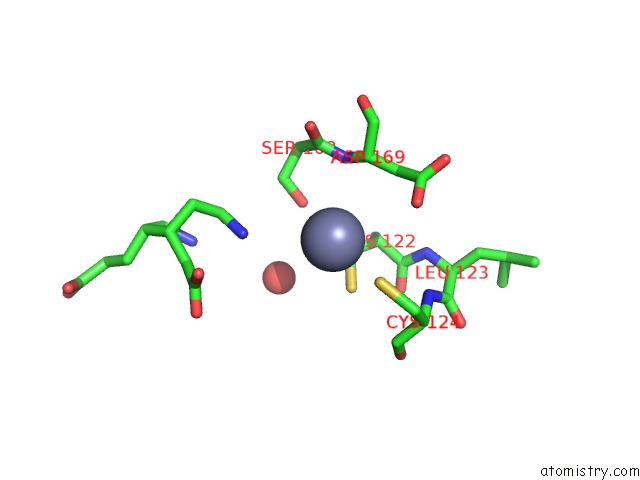
Mono view
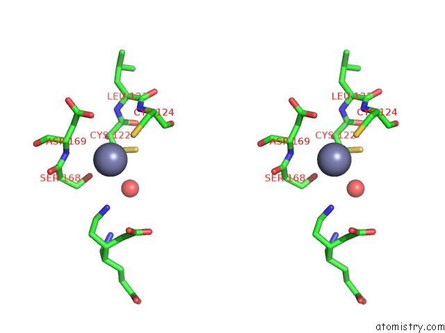
Stereo pair view
Mono view
Stereo pair view
A full contact list of Zinc with other atoms in the Zn binding
site number 1 of Crystal Structure of A Low Activity F12L Mutant of Human Porphobilinogen Synthase within 5.0Å range:
|
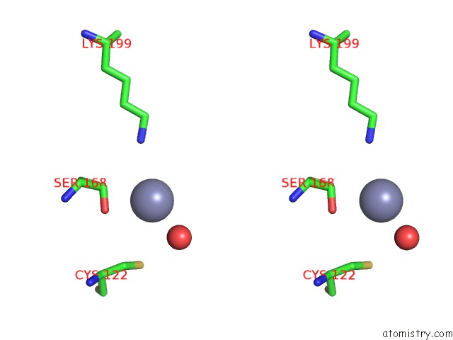
Zinc binding site 2 out of 2 in 1pv8
Go back to
Zinc binding site 2 out
of 2 in the Crystal Structure of A Low Activity F12L Mutant of Human Porphobilinogen Synthase
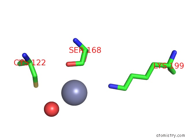
Mono view
Stereo pair view
Mono view
Stereo pair view
A full contact list of Zinc with other atoms in the Zn binding
site number 2 of Crystal Structure of A Low Activity F12L Mutant of Human Porphobilinogen Synthase within 5.0Å range:
|
Reference:
S.Breinig,
J.Kervinen,
L.Stith,
A.S.Wasson,
R.Fairman,
A.Wlodawer,
A.Zdanov,
E.K.Jaffe.
Control of Tetrapyrrole Biosynthesis By Alternate Quaternary Forms of Porphobilinogen Synthase. Nat.Struct.Biol. V. 10 757 2003.
ISSN: ISSN 1072-8368
PubMed: 12897770
DOI: 10.1038/NSB963
Page generated: Wed Oct 16 17:56:34 2024
ISSN: ISSN 1072-8368
PubMed: 12897770
DOI: 10.1038/NSB963
Last articles
Zn in 9J0NZn in 9J0O
Zn in 9J0P
Zn in 9FJX
Zn in 9EKB
Zn in 9C0F
Zn in 9CAH
Zn in 9CH0
Zn in 9CH3
Zn in 9CH1